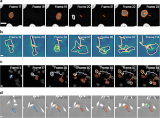
MAGIK reliably links trajectories in
various experimental scenarios. a, Confocal microscopy of green fluorescent
protein (GFP)-transfected GOWT1 mouse stem cells. MAGIK achieves an F1 score of 99.8% and TRA = 99.2% despite the
fact that the cells frequently leave the field of observation. Scale bar,
10 μm. b, Phase-contrast imaging of glioblastoma-astrocytoma U373 cells on a
polyacrylamide substrate. MAGIK reaches an F1 score of 99.8% and TRA = 100% even though
the cells greatly change shape over time. Scale bar, 10 μm. c, Epifluorescence
imaging of HeLa cells stably expressing histone H2b–GFP. MAGIK achieves an F1 score of 98.8% and TRA = 98.4% despite the
dense sample and frequent mitosis and collisions. Scale bar, 10 μm. d,
Phase-contrast imaging of pancreatic stem cells on a polystyrene substrate.
MAGIK obtains an F1 score of 99.3% and TRA = 98.5% despite high cell
density, elongated shapes, pronounced cell displacements and a significant
number of division events. Scale bar, 10 μm. Interrupted trajectories
correspond to cases where cells left the field of view or missed segmentation
in the image sequence. All videos belong to the dataset of the 6th Cell
Tracking Challenge. Credit: Nature Machine Intelligence (2023). DOI: 10.1038/s42256-022-00595-0
The
enormous amount of data obtained by filming biological processes using a
microscope has previously been an obstacle for analyses. Using artificial
intelligence (AI), researchers at the University of Gothenburg can now follow
cell movement across time and space. The method could be very helpful for
developing more effective cancer medications.
Studying the movements and behaviors of
cells and biological molecules under a microscope provides fundamental
information for better understanding processes pertaining to our health.
Studies of how cells behave in different scenarios is important for developing
new medical technologies and treatments.
"In the past two decades, optical microscopy has advanced significantly. It enables us to study biological life down to the smallest detail in both space and time. Living systems move in every possible direction and at different speeds," says Jesús Pineda, doctoral student at the University of Gothenburg and first author of the scientific article in Nature Machine Intelligence.
A. Linking
of HeLa cells. MAGIK successfully tracks (TRA = 99.2%) HeLa cells on a flat
glass substrate despite the changes in shape, the high packing density, the low
SNR and the heterogeneous dynamics as a consequence of their migration and
proliferation. B. Linking of pancreatic stem cells. MAGIK successfully tracks
pancreatic stem cells on a polystyrene substrate despite the high cell density,
the elongated shapes, the pronounced cell displacements and a significant
number of division events (TRA = 98.5%). Credit: Jesús Pineda et al,
Mathematics describes relationships of
particles
Advancements have given today's researchers such large amounts of data that
analysis is nearly impossible. But now, researchers at the University of
Gothenburg have developed an AI method combining graph theory and neural networks that can pick
out reliable information from video clips.
Graph theory is a mathematical structure that is used to describe the
relationships between different particles in the studied sample. It is
comparable to a social network in which the particles interact and influence
one another's behavior directly or indirectly.
"The AI method uses the information in the graph to adapt to different
situations and can solve multiple tasks in different experiments. For example,
our AI can reconstruct the path that individual cells or molecules take when
moving to achieve a certain biological function. This means that researchers can
test the effectiveness of different medications and see how well they work as
potential cancer treatments," says Jesús Pineda.
AI also makes it possible to describe all dynamic aspects of particles in situations where other methods would not be effective. For this reason, pharmaceutical companies have already incorporated this method into their research and development process.
Source: AI analyzes cell movement under the microscope (phys.org)

No comments:
Post a Comment